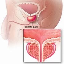
The prostate is a male-specific gonadal organ. The prostate is a pair of parenchymal uterus composed of glandular tissue and muscle tissue. Prostate looks like chestnut, is in the front of the pubic symphysis and back of the rectum. There is a urethral passage in the middle of the prostate gland, holding the urethral orifice. Therefore, the urination is affected first by prostate disease. Prostate is a rare sexual secretory gland with both endocrine and exocrine functions.
As an exocrine gland, the prostate secretes about 2 milliliters of prostatic fluid every day, which is the main component of semen. As an endocrine gland, the hormone secreted by the prostate is called "prostaglandin".
The normal prostate is 4 cm in diameter, 3 cm in diameter, 2 cm in diameter and 20 g in weight. It consists of 30-50 alveolar glands with 15-30 excretory ducts opening on both sides of the spermatorrhea. The size and weight of the prostate varied with age, increased with the development of puberty, peaked at the age of 24, and gradually degenerated and atrophied after entering old age.

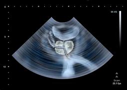
Prostate ultrasonography is a ultrasound imaging technique for the prostate. The size, shape, capsule and blood flow of the prostate can be observed by prostatet ultrasond report, and abnormal conditions of gland lesions and stones can be detected. Prostate is a specific gonadal organ for male, and the daily secretion of prostatic fluid is the main component of semen, which plays an important role in fertility. The prostate wraps around the urethra and holds the urethral orifice, affecting urination. Once prostate disease occurs, it will affect urination and reproductive function.
Normal analysis of ultrasound reports of prostate : the prostate is chestnut-shaped, the internal echo is uniform low echo type, and the internal and external glands of thin people are clearly demarcated. The transverse diameter is less than 4 cm, the upper and lower diameter is less than 3 cm, the anterior and posterior diameter is less than 2 cm, and the volume is generally less than 20 ml.

Abnormal analysis of ultrasound reports of prostate: prostate hyperplasia is characterized by enlargement of prostate volume, obvious enlargement of internal gland, moderate or medium-strong echo, uneven internal echo, and often calcification. The enlarged prostatic gland often protrudes into the bladder with abundant blood flow, which can be seen around the proliferative nodules.
prostatitis is partly manifested by flake-like lesions in the gland below the echo area and dilatation of the periprostatic venous plexus in the previous case; when accompanied with abscess, the volume of the prostate enlarges, while hypoechoic lesions and irregular shapes appear in both internal and external glands,the liquid dark areas are visible in it; and blood flow information increases.
At present, prostate ultrasonography can not reflect the histological and cytopathological characteristics of prostate. Therefore, we must make a diagnose combining the ultrasound report with the anatomy, pathology and clinical knowledge , so as to make correct conclusions.

